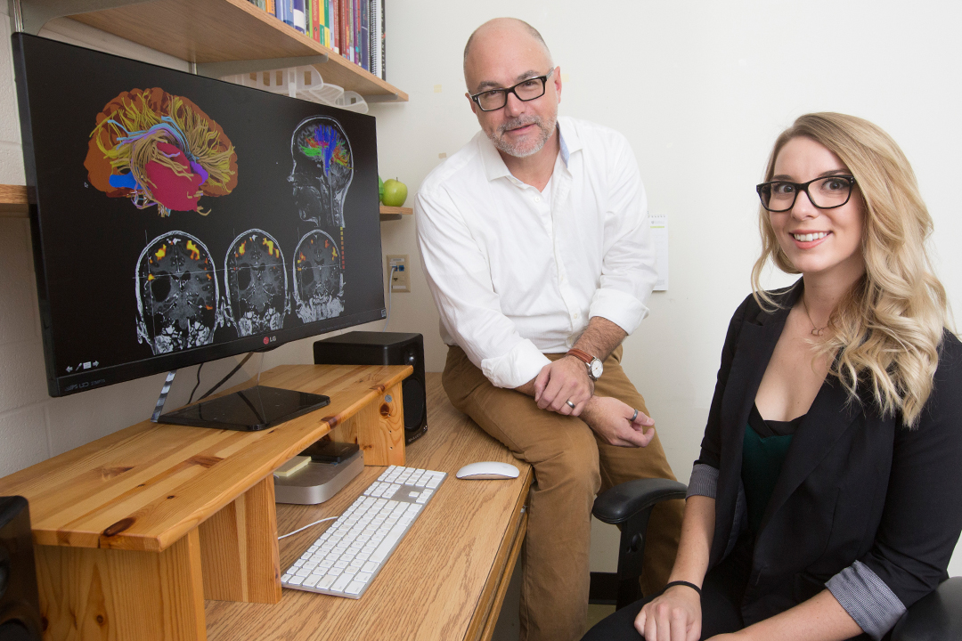
Working with cognitive neuroscience professor Ron Borowsky, she is combining two well-known imaging techniques with novel 3D mapping to look at cognitive areas of the brain in people about to undergo surgery.
This new approach, developed during Ekstrand’s recently earned master’s degree, is already helping Saskatoon surgeons plan brain surgeries by providing information about brain areas to avoid during surgery. This information is not available from regular scans such as computed tomography (CT) or magnetic resonance imaging (MRI).
“Our team has collaborated with neurosurgeons in over two dozen cases,” said Borowsky. “This has resulted in important advances in pre-surgical planning in Saskatoon, enabling even safer and more effective neurosurgeries.”
Ekstrand creates super-detailed brain maps of patients at Royal University Hospital in Saskatoon using innovative 3D visualization software. These maps combine information from two widely used techniques ‒ functional magnetic resonance (fMRI) and diffusion tensor imaging (DTI).
One example is Ekstrand’s 3D brain scan of a 44-year-old woman whose motor and sensory areas in the brain had shifted to surround a large tumor. The surgeon removed the tumor by planning a surgery that avoided cutting areas that could have caused permanent impairment. Instead, the surgery resulted in only minor temporary side effects.
“In Canada there aren’t many centers performing pre-surgical fMRI and DTI in 3D,” said Ekstrand. “This new 3D approach could be extremely valuable to neurosurgeons.”
Ekstrand looks at how parts of grey matter in the brain activate when patients touch objects, and how this relates to the relatively uncharted connection between memory and the sense of touch.
“The brain has always fascinated me,” she said. “This tiny organ orchestrates our entire lives, defines who we are, and yet we know virtually nothing about it.”
She used fMRI on a patient’s brain asking her to perform tasks such as squeezing a stress ball with both hands, licking her lips, and rubbing her feet together to understand which parts of the brain control these movements.
She also used the DTI to visualize how the touch sensory area connects with other brain areas through white matter tracts, the network of neural “wires” enabling communication among brain areas.
Early fMRI results using a task that asks participants to say how they would interact with a pictured object suggest that touch plays a role in how we remember to use objects. But further research is needed, said Ekstrand.
Helped by engineering student Devin Bradburn, Ekstrand also mapped and printed 3D models of her own brain, thus creating a tangible and anatomically correct human brain. U of S neurosurgery professor Ivar Mendez now uses these models for teaching surgery and for experimenting on the implantation of stem cells for patients with Parkinson’s disease.
Through this work funded by the federal agency NSERC, Ekstrand has become the first author in a publication for Frontiers in Integrative Neuroscience. She will focus her PhD project on the brains of epileptic patients.
Federica Giannelli is a graduate student intern in the U of S research profile and impact unit.
This article first ran as part of the 2016 Young Innovators series, an initiative of the U of S Research Profile and Impact office in partnership with the Saskatoon StarPhoenix.