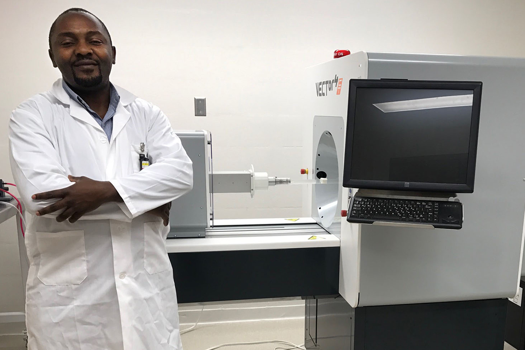
The scanner—a microPET/ SPECT/CT—is the first big building block in the new medical imaging research lab that will help to do research development of radiopharmaceuticals—pharmaceuticals labeled with isotopes that are used for diagnosis and therapy—at the U of S.
“The first phase of this program, which was funded by the provincial and federal governments, allowed the university to establish the Saskatchewan Centre for Cyclotron Sciences, which is currently being used to produce agents or drugs that are already approved,” said Humphrey Fonge, a clinical assistant professor in the department of medical imaging. “That kind of facility exists in almost every province in Canada.” But the new Preclinical Imaging Facility and Radiopharmaceutical Lab takes that work one step further by giving researchers in the College of Medicine the opportunity to design, and ultimately produce, their own drugs.
“Existing compounds that are currently used to diagnose patients (can be) kind of sub-optimal. In some cases, they don’t give the best diagnostic characteristic of the patients, and there are very few of such compounds that are used by Health Canada,” Fonge said, explaining the importance of continuing research into pharmaceutical production. “So what that means because it’s not a one-size-fits-all, physicians in certain cases cannot make the right diagnosis based on the existing diagnostic tools that they have.
“So the research in this area is very intensive, and very rapidly growing, whereby we seek to develop newer agents that would (help) physicians improve diagnosis, compared to what they currently have.”
The need to advance that research is what led the team to this second phase of the program, which was the creation of this lab.
“It’s going to be designated for anyone, like a core facility,” Fonge explained. “Anyone whose research interest is to develop new diagnostic agents for molecular imaging. It’s similar to the concept in health sciences: shared labs.”
The work starts in the radiochemistry lab, where the researchers work on designing and producing the initial radiolabeled drugs for testing. After in vitro testing, they move on to the invivo studies, and that’s where the new scanner comes into play. Following injection of the radiolabeled drug in a living subject, the microPET/SPECT/CT picks up radiation from the subject which is being scanned, allowing a computer to stitch together the resulting images in a process called ‘construction.’
The end result is a composite that allows the researcher to understand the structural and molecular information of the organs, giving them the ability to clearly characterize the diseased organs/tissue.
“The nice thing about this kind of work is how interdisciplinary it is,” Fonge said. “I know on campus there are four principal investigators (PI) whose research is solely this, and three of them have just been hired in the last three months. There aren’t many universities in Canada that have that number of really high profile PI’s in this field, so the university being able to attract people like that is a good thing.”
The lab, which officially opened in mid-December, hasn’t finished growing quite yet. Fonge and his team—with support from the university—recently submitted an application for an $18 million Canada Foundation for Innovation (CFI) grant and will find out in June if they have been successful.
Marg Sheridan is an online communications co-ordinator in the College of Medicine.