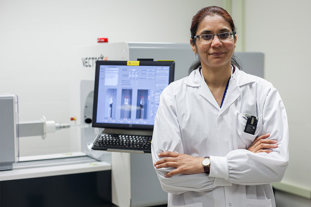
What happens inside these spongy, air-filled organs may be key to unravelling the mysteries of a wide array of illnesses.
“I think by studying lungs I can study a lot of other organ systems … It will keep me motivated over the years,” Aulakh said, an assistant professor in the Western College of Veterinary Medicine’s (WCVM) Department of Small Animal Clinical Sciences.
That line of inquiry comprises much of her work as the University of Saskatchewan’s first Fedoruk Chair in Animal Imaging, a position supported by the Sylvia Fedoruk Canadian Centre for Nuclear Innovation.
Her path leading to this point began in the Indian state of Punjab where she grew up. The daughter of two teachers and a self-described “geek,” she declared in a childhood essay that she wanted to be a research scientist.
Actually, her first choice was to follow in the footsteps of her grandfather, an anesthesiologist in the Indian army.
“He was a doctor with great stories to tell as a saviour of lives,” she recalled.
But when Aulakh’s medical application was unsuccessful she turned to pharmacy, confident it could lead to a research career. After completing her master’s degree in pharmacology at Punjabi University, Aulakh took a job as a research scientist and became heavily involved in a project dealing with lung inflammation. Determined to pursue higher studies, she sought an opportunity to keep working on that topic. When former WCVM professor Baljit Singh recruited her to the U of S, “that was the eureka moment,” Aulakh said.
It seemed that everything was coming together for her. As she flew into Saskatoon on a September day in 2007, Aulakh saw wheat fields just like back home in Punjab, India’s own bread-basket. Singh, whoservedasher research mentor, was also educated in Punjab and his own research focused on lung inflammation.
“It gave me some reference point, context for my past research. And it really gave me motivation to pursue that direction,” Aulakh said.
She also credits Singh for her crossover to work in medical imaging. It happened when Aulakh was studying one particular protein molecule called angiostatin. Since there were no tools on the market that would allow her to look at angiostatin and see what it does, the research team worked to “label” the protein molecule in their own lab (chemically attaching a label or fluorescent tag to aid in the molecule’s detection).
The molecule was “mesmerizing” to look at, Aulakh said. “It was so surreal. I mean, you look at what it was doing to a cell.”
The WCVM research team moved on to the problem of imaging lungs. Because lungs contain a lot of air, they are “basically not visible” and are elusive subjects, explained Aulakh. Singh and Aulakh discussed the problem with Dean Chapman, now science director at the Canadian Light Source (CLS) at the U of S. She describes Chapman as a “master of technique” in a type of X-ray microscopy called multiple image radiography, and said he uses a novel stabilized setup for this at the CLS.
“We were convinced that this technique would look at those air-tissue interfaces in a way that no other technique can,” she said.
Their findings led to further work that focused on ways to see lung inflammation. Singh, Chapman and Aulakh collaborated with Dr. Wolfgang Kuebler—a renowned lung scientist at the University of Toronto.
Aulakh is intrigued by diseases that seem to be unrelated to the lungs—but still turn out to have surprising effects on these organs. For instance, an equine disease called laminitis that severely affects horses’ feet also causes issues in their lungs.
“Why in the world would this happen?” said Aulakh.
Similarly, dogs with immune-mediated hemolytic anemia have heavily-damaged lungs because they have been infiltrated with a host of immune cells.
Aulakh believes the lungs’ ability to act as reservoirs of neutrophils may begin to explain why they too are damaged by diseases that target other parts of the body.
In Canada, there is likely no better place than the U of S for a scientist who approaches the study of disease by following the path of one molecule through an animal. With the combined facilities of the CLS and the Saskatchewan Centre for Cyclotron Sciences, the U of S is a one-stop shop.
Indeed, Aulakh said the dual PET-SPECT scanner at the cyclotron can image an entire mouse while the CLS’s biomedical beamline can image anything from the size of a fly to a horse.
“We are really pushing ourselves to produce some groundwork in the disease models that we have ventured upon for these next couple of years,” she said.
Kathy Fitzpatrick is a freelance journalist in Saskatoon. Born in Manitoba, she has spent close to four decades working in media.
