
Researchers explore novel prostate cancer imaging techniques
A team of U of S researchers is developing a new window into a deadly disease that could ultimately help not only men, but also man’s best friend.
By Michael Robin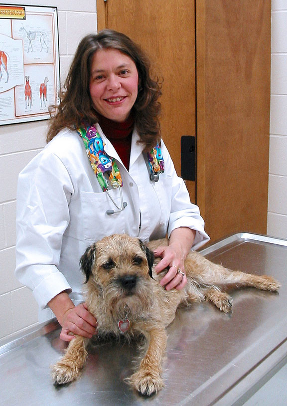 "We are looking at prostate cancer in dogs as a model for human disease," said Murray Pettitt, a researcher with the Department of Animal and Poultry Science in the College of Agriculture and Bioresources. "Dogs are the only other large mammals that have a significant incidence rate of spontaneous prostate cancer."
The U of S Prostate Research Team is working at the Canadian Light Source (CLS) to make the process of diagnosing prostate cancer more accurate and less invasive. The nine-member team draws expertise from the Colleges of Medicine, Veterinary Medicine and Agriculture and Bioresources, as well as the Saskatchewan Cancer Centre and the Saskatoon Health Region.
Physicians have several tools at their disposal for diagnosing prostate cancer, including blood tests and imaging technologies, but to get a definitive diagnosis, doctors must collect a biopsy. The team hopes to change this by coming up with a way to produce images of the prostate with enough detail to either diagnose cancer directly, or at least pinpoint the areas of interest for later biopsy. Since it had never been done before, the team found their ingenuity pushed to new limits.
"You don't just go to the synchrotron and do research," said Dr. Liz Snead, a small animal internal medicine specialist at the Western College of Veterinary Medicine (WCVM). "I honestly feel like McGyver every time we go there because you have to work it all out yourself. While the CLS is extremely supportive in getting you on the beamline and providing the basic set up for the type of imaging you want to do, these experiments by their nature are breaking new ground. That means you have to tailor the setup precisely to get your image."
This means a lot of trial and error – from figuring out how to set up a prostate in a jar to image it to manufacturing a cradle to hold a dog in the beam for a non-invasive scan. Pettitt said the learning curve has been steep but a technique called phase contrast CT imaging provided the image quality they wanted.
"It was Christmas that day, let me tell you," Pettitt said. Snead agreed.
"I think we've all been blown away at what we're getting from the phase contrast imaging," she said. "When we first saw the image, our eyes just went really wide. We were all like, ‘wow!' – it was really impressive."
The phase contrast images show so much detail the team compares it to viewing tissue on a microscope slide.
"You can see individual glands and the ducts where the glands feed into the urethra," said Dr. James Montgomery, a medical imaging specialist with the WCVM.
"There's still stuff we need to work out to go from looking at a prostate out of a body to looking at it in a live animal or person, but that's where we're headed."
The ultimate goal, Pettitt said, is creating a detailed 3D image of a patient's prostate right on a clinician's computer screen, an image that could be manipulated on screen and peeled away layer by layer to examine the gland in detail.
The team has made important first steps, thanks to their unique collaboration and close access to the CLS. Funding support from Canada's Motorcycle Ride for Dad, the Saskatchewan Health Research Foundation, and the Sylvia Fedoruk Canadian Centre for Nuclear Innovation plus three U of S colleges has also been essential.
While much of the attention has focused on prostate cancer in humans, the research will also benefit dogs. Snead explained that dogs cannot tell their owners early if they're feeling bad "down there." Consequently, by the time the cancer is diagnosed, the veterinarian usually has one option: palliative care. An imaging tool could give vets a chance to catch it in time to treat it.
"I don't know if we've even scratched the surface of what is potentially possible with this type of imaging," Snead said.