
Bones of contention
In Canada, bone fractures due to osteoporosis affect one in three women and one in five men over their lifetimes, costing the health-care system more than $2.3 billion a year.
By University Communications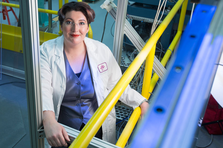 But despite the high incidence of this devastating disease, scientists know little about its causes and why the disease disproportionately affects women.
At the University of Saskatchewan, a team led by Canada Research Chair David Cooper aims to better understand how bones age and what differences exist between the sexes.
"This research could lead to new targets for maintaining bone health over the lifespan, halting bone loss and, ideally, restoring compromised bone," says Cooper.
Recent PhD graduate Yasmin Carter is exploring why the disease attacks women's bone structure. She is looking at small cavities in bone that contain cells regulating how much bone a person has.
"I have identified, for the first time, a possible difference in structure between the bones of men and women," said Carter.
Her research, recently published in the Journal of Structural Biology, is the first large-scale study to show that unlike in men, bone cell spaces in women start off larger and become smaller with age, possibly preventing cells from functioning properly.
Previous studies had connected bone cell death to a decline in the number of cells, a possible cause for osteoporosis.
"Yasmin has found that while the numbers of bone cells do not change in women with age—as some have suggested—the size and shape of cellular spaces do. Briefly, the spaces get smaller and rounder in shape," said Cooper.
These cavities are linked through a network of channels that, similar to a plumbing system, regulate exchange of nutrients, wastes and bone regeneration. She was able to prove that these cavities and their communication system become smaller with aging in women, possibly due to the requirements of pregnancy.
Carter suspects a process known as infilling causes this change. Bone cavities may get smaller as minerals progressively form a plug on their surface, impeding their regular functions and causing cell death over the long term.
Infilling may also damage the cell networking that regulates bone turnover, the process of removing old bone and replacing it with healthy bone.
"This process may explain the decrease in bone material strength with aging, making it more susceptible to fractures in the long-term," said Carter.
As bone cavities are not detectable using regular medical X-ray devices, Carter collected data at an American synchrotron, scanning women's bone samples and measuring cell cavities in females aged 20 to 86.
Hers is the first large-scale study to use 3-D computed tomography (CT) scannning to visualize bone cell spaces. The bone samples came from one of the best bone tissue collections in the world—the Melbourne Femur Collection at the University of Melbourne.
Cooper is commissioning new equipment to do this type of research at the Canadian Light Source (CLS) synchrotron at the U of S. Carter used the CLS as a training ground to learn techniques and gain first-hand experience she later applied to her research.
Her preliminary research paves the way for further studies on the link between osteoporosis and infilling, and possibly for development of new drugs.
The research was funded by the U of S, Saskatchewan Health Research Foundation, and a federal training program in health research using synchrotron techniques. Carter's work is featured in a video at: http://www.usask.ca/research/communications/multimedia/ideas.phpArticle written by Federica Giannelli, a graduate student intern in the U of S research profile and impact unit. This article first ran as part of the 2014 Young Innovators series, an initiative of the U of S Research Profile office in partnership with the Saskatoon StarPhoenix.