
Improved “camera pill” tech attracts funding for engineering researcher
Electrical and computer engineering associate professor Khan Wahid has been awarded a further $99,500 boost to help develop new wireless video software and hardware for endoscopy capsules, a sort of electronic “camera pill,” promising doctors a much-improved window into the inner workings of their patients.
By Michael Robin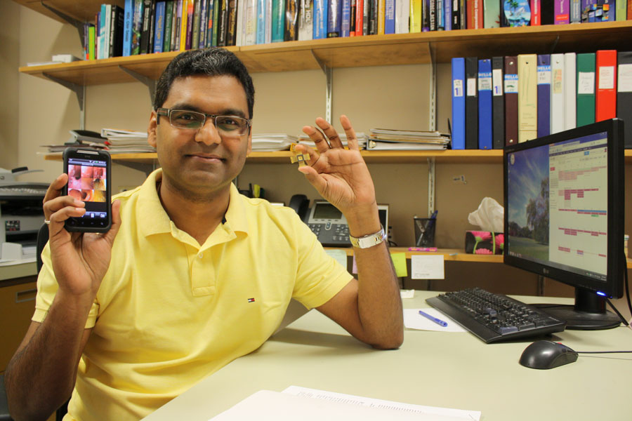 "Doctors are not satisfied with the current image quality from endoscopy capsules. We are working to improve the technology in several ways, which will lead to more consistent, accurate diagnosis," Wahid said.
The latest funds, provided through Western Economic Diversification Canada, add to resources previously provided through Grand Challenges Canada and the Canada Foundation for Innovation to build a leading-edge research facility in the College of Engineering for Wahid to further develop and commercialize his next generation endoscopy system.
Endoscopy capsules contain a video camera, battery, light-emitting diode (LED) and a transmitter, all packed into a package about the size of a large vitamin pill. Once swallowed, they transmit pictures of a patient's inner workings, providing doctors with invaluable information for diagnosing ailments such as causes of internal bleeding or abdominal pain such as Crohn's disease, peptic ulcers or colorectal cancer.
Wahid's patent-pending solution has overcome two major disadvantages of existing capsules: the limited number of pictures due to battery life, and the bulky data capture device patients must wear.
"Our technology offers significant improvements over existing systems in terms of comfort for the patient, but more importantly, a much richer information stream that will allow doctors to make faster and better diagnoses," Wahid said.
His novel wireless image compression software and custom microchip solution offers superior images at increased frame rates and a new feature: multi-spectral imaging. Much like coloured filters enhance certain elements in photographs, multi-spectral imaging allows doctors to choose which version of an image best highlights the tissue they are looking at. The bottom line is more and better information with which to make a diagnosis. Data from the capsule can be transmitted to the patient's smart device such as a cell phone or transmitted to a hospital network via Wi-Fi or internet to enable real-time and remote diagnosis.
Wahid and his team will now put the technology through extensive testing and prototyping before taking the next steps to human trials, aided by the U of S Industry Liaison Office. This may include establishing a local spin-off company to manufacture the add-on chips and market the technology to the international medical community, or a joint venture with an existing medical device company.
Wahid explained the versatile technology may eventually find uses in the industrial, security and environmental sectors, but for now, the focus is medicine.
"We're taking it one step at a time."