
U of S imaging technique advancing stroke treatment
A new University of Saskatchewan technology combining innovative synchrotron imaging and 3D printing could be a game changer for more accurate stroke prediction and for ultimately guiding surgery.
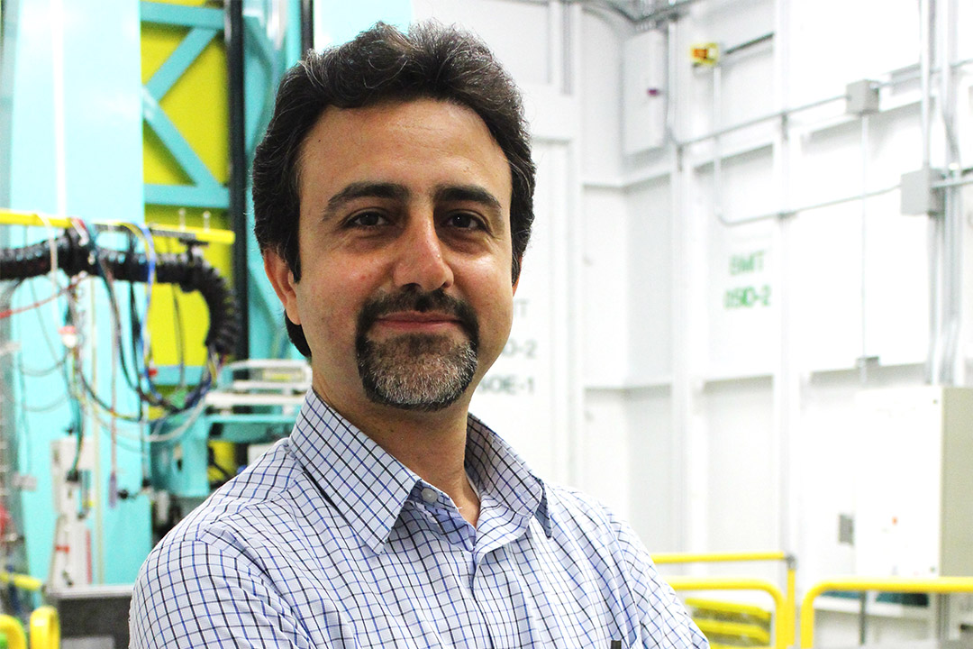
By Federica GiannelliIn a journal article published this week in Biomedical Physics & Engineering Express, post-doctoral fellow Mohammad Izadifar’s preliminary results show that unlike regular medical imaging, his new “blood-flow mapping technique” can determine whether a patient’s aneurysm, a tiny blood balloon in a brain artery, is about to burst causing bleeding (hemorrhagic stroke).
“When a patient shows up with an aneurysm at the hospital, doctors usually don’t know when or whether it will burst,” said Izadifar, originally from Iran and now a Canadian citizen.
The 60,000 new stroke cases in Canada each year cost the healthcare system over $3.6 billion yearly. Strokes happen because of blood clots in a brain artery (ischemic strokes), but up to 20 per cent of aneurysms may burst and bleed, causing hemorrhagic strokes.
“With regular medical imaging, it is very difficult for clinicians to tell the difference between stable aneurysms that could undergo surgery and those likely to rupture,” said Izadifar.
He said that with his synchrotron technique he was able to identify specific areas of the aneurysm that could burst.
“Our new technique could provide neurosurgeons with a new avenue for pre-assessment of different treatment options,” said U of S surgery professor Lissa Peeling, Izadifar’s collaborator along with surgery professor Michael Kelly.
Izadifar designed a 3D-printed model of a patient’s aneurysm based on the patient’s MRI brain scan. At the U of S Canadian Light Source synchrotron, he connected that model to a blood circulation system that simulates a human heartbeat. Using his new imaging technique, he was able to visualize the blood flow patterns and pressure in the 3D model.
“I was able to track the footprint of blood cells in the flow to see where they are going in the aneurysm,” said Izadifar, who has received funding from the Saskatchewan Health Research Foundation (SHRF) and the Society of NeuroInterventional Surgery (SNIS) Foundation.
At the Royal University Hospital, Izadifar tested the 3D aneurysm model using CT scanning with a contrast agent, but he couldn’t track the blood flow. So he assessed the potential of using the powerful X-ray synchrotron light and it worked.
Unlike MRI and X-ray scans, Kelly said the new imaging technique is remarkably superior because it works without contrast agents that chemically enhance the visibility of blood vessels in the body.
While his imaging takes under five minutes, Izadifar said more research is needed to develop new software able to reconstruct the data collected fast enough for potential clinical applications in stroke treatment.
“Now I want to test this technique on a larger scale on aneurysms of different size and shape to simulate real-life situations,” he said.
His preliminary study also shows that stents, minimally invasive devices inserted into brain arteries, could prevent strokes with a blood flow reduction of 50 to 70 per cent in aneurysms. But Izadifar cautions more research is needed to confirm his initial results.
Federica Giannelli is a graduate student intern in the U of S research profile and impact unit.
This article first ran as part of the 2017 Young Innovators series, an initiative of the U of S Research Profile and Impact office in partnership with the Saskatoon StarPhoenix.