
‘Deep learning’ software automatically detects diseases
Patients could soon get faster and more accurate diagnoses with new software that can automatically detect signs of diabetes, heart disease and cancer from medical images.
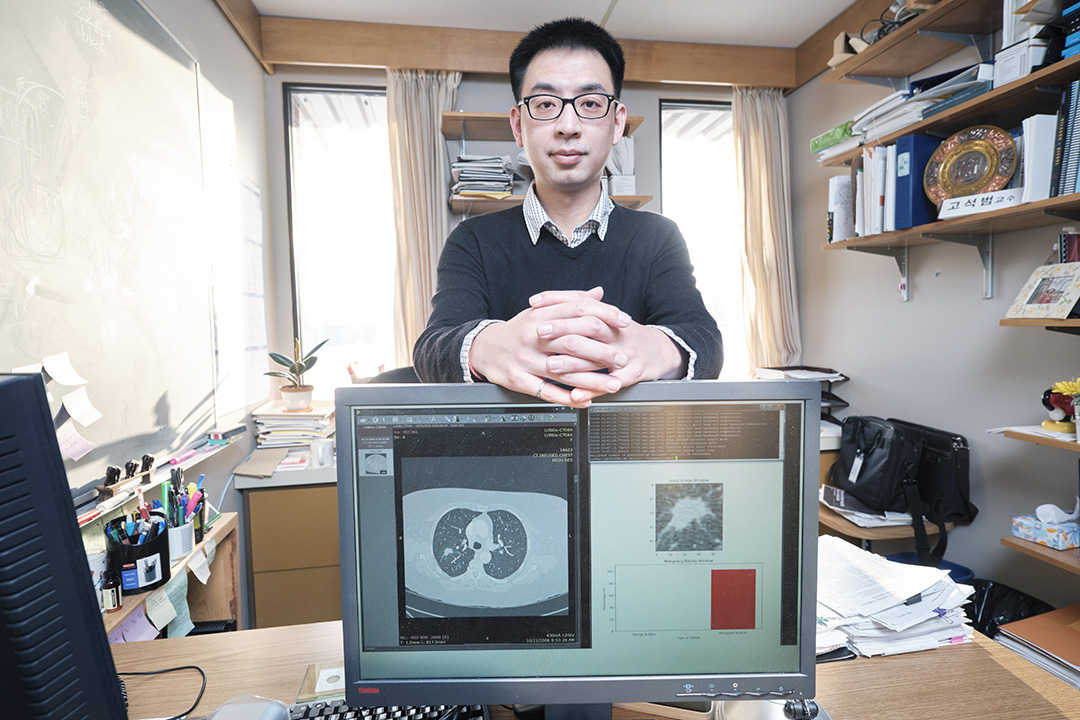
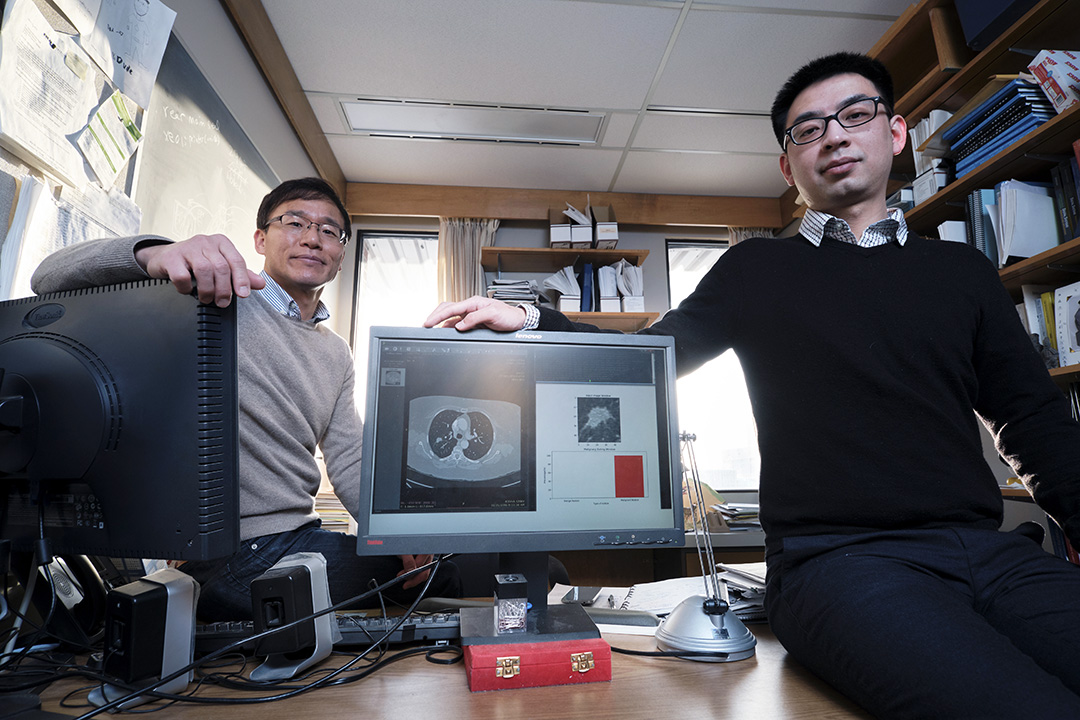
By Federica GiannelliUniversity of Saskatchewan PhD student Yi Wang developed software that can get higher image quality. It improves current computer-aided diagnosis (CADx) technology, which assists doctors to detect diseases from medical imaging scans such as ultrasound, computer tomography (CT) and retinal fundus imaging, which captures photos of the back of the eye.
Wang’s software makes diagnosis faster — it takes less than 30 seconds and it is around 10 times faster than current ones.
“Our software will help medical staff reduce the time they take to interpret medical images, so that they can provide better patient care,” said Seok-Bum Ko, an electrical and computer engineering professor and Wang’s supervisor. “Radiologists and doctors can use their saved time more efficiently for other important tasks.”
Wang has tested his software on detecting abnormal retinal blood vessels in the eye — a symptom common to diabetes or heart disease — and was 97 per cent accurate at identifying abnormal vessels that needed further diagnosis. The results are published in the journal of Computerized and Medical Imaging Graphics.
Detecting blood vessels from retinal fundus imaging is often difficult. The images may end up blurred, so the vessels may be difficult to identify. Also, doctors usually have to mark blood vessel patterns manually on the image to determine whether vessels are broken — a time consuming process.
Wang’s software uses a state-of-the-art system called “deep learning” that helps improve image classification and quality.
“Deep learning relies on software algorithms that make the software automatically learn and analyze image patterns,” said Wang, a student from China. “The idea is that the more images the software ‘reads,’ the better and more accurate it becomes at distinguishing healthy vessels from broken ones, so we may say it progressively ‘learns.’ This idea is at the core of all studies on artificial intelligence.”
To prove that his software works, Wang tested it on more than 130 images taken from a public database where diagnoses were already available, so that he could compare systems. His software is proved to be two per cent more accurate than commercial counterparts.
“Our software is a good tool to complement radiologists’ and doctors’ expertise, not to substitute it,” said Ko. “There is a concern that this type of new ‘intelligent’ technologies will replace humans, like in science fiction. That is not the case, because we will always need people to make machines work.”
Wang and Ko, who have been awarded funding from the federal agency NSERC, are already teaching the software to detect lung and breast cancer from CT and ultrasound images respectively, with very positive results.
“We are very excited about our detection system, and we are sure it will also make a change in medical teaching,” said Ko.
Future applications may include using the software to teach medical students how to recognize diseases from CT images. Ko tested it on researchers at Chonbuk National University, South Korea, in a class setting and said early results are very promising.
This article first ran as part of the 2019 Young Innovators series, an initiative of the U of S Research Profile and Impact office in partnership with the Saskatoon StarPhoenix.