
Bacteria on the surface of the eye not all the same: new insights from USask research
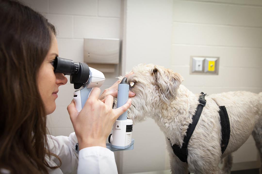
A pioneering study led by University of Saskatchewan (USask) veterinary ophthalmologist Dr. Marina Leis (DVM, DACVO) shows that bacterial communities vary on different parts of the eye surface—a finding that significantly alters understanding of the mechanisms of eye disease and can lead to developing new treatments.
“We are excited to share our findings, which provide a paradigm shift within the field,” said veterinary microbiologist Dr. Matheus Costa (DVM, PhD), a member of the Leis research team that published a paper recently (Feb. 19) in the peer-reviewed scientific journal PLOS One.
Team members are Costa and Leis, assistant professors at the Western College of Veterinary Medicine (WCVM) at USask, and Gabriela Madruga, a PhD student from São Paulo State University in Brazil.
The pilot study was conducted on eyes from 15 piglets euthanized for reasons unrelated to this research. Piglets were chosen as models because pigs’ eyes are relatively similar to human eyes. This model allowed researchers to sample the cornea without use of topical anesthetic drugs that might disturb the microbiota of the eye.
Researchers found that the corneal surface provides a distinct environmental niche within the ocular (eye) surface, leading to a bacterial community different from all other sample types.
“The way we’ve always understood the ocular surface was that it contained a single bacterial population. Now, we learned different portions of the surface seem to have different bacteria that predominate, which has implications for disease mechanisms of multiple types of ocular surface conditions,” Costa said.
“As ophthalmologists we work under the assumption that the cornea is largely devoid of bacteria, or at least clinically relevant players. What we found puts this view into question,” Leis said.
For example, two distinct types of bacteria were found in a significantly larger proportion in association with the cornea.
“These two types of bacteria not only have a reported association with the ocular surface in the literature but also have the potential to cause opportunistic infection,” Leis said.
The findings demonstrate that the location on the eye surface matters during routine collection of a swab for diagnostic purposes, Leis said.
Leis has secured funding for follow-up translational studies targeting other species of interest to veterinarians, especially companion dogs that are susceptible to dry eye disease—a very common chronic condition that results in discomfort and sometimes even blindness.
Translation of the findings to human health likely is a long way off, until the results are further validated through research involving smaller animals such as dogs and larger animals such as horses.
The study was conducted with funding from the WCVM Companion Animal Health Fund and the Department of Veterinary Clinical Sciences at the University of Minnesota.