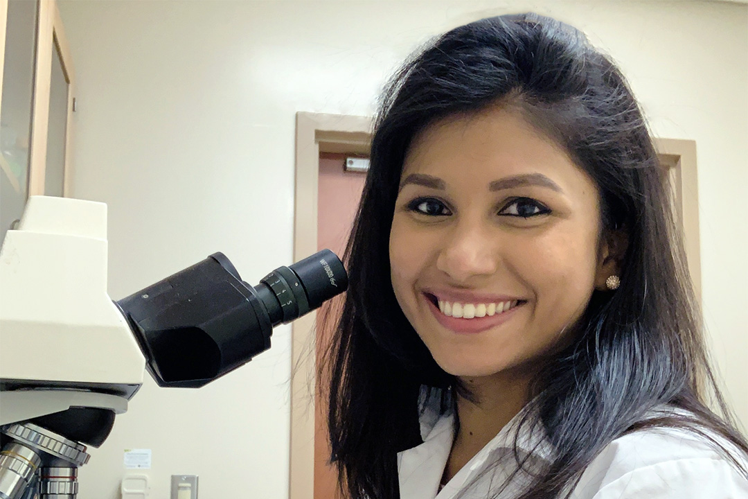
USask researcher uses innovative imaging techniques to determine blood clot composition
An innovative new project by a U of S graduate student used synchrotron-based imaging techniques to examine what blood clots are made of.
By BROOKE KLEIBOERStrokes are the third-leading cause of death in Canada. According to the Canadian Medical Association, every 10 minutes a Canadian suffers a stroke.
Treating them effectively is vitally important, and is necessary to address the post-immunization appearance of blood clots that has become prevalent during the COVID-19 pandemic.
Presently, 80 per cent of Canadians will survive a stroke event with timely treatment, but they often end up with mild to severe permanent disability. University of Saskatchewan cerebrovascular research aims to improve these statistics.
An innovative new project by a U of S graduate student used synchrotron-based imaging techniques to examine what blood clots are made of, and how composition could potentially affect stroke treatment options and patient outcomes. According to the Heart and Stroke Foundation of Canada, the recovery rates are 70 per cent, but the team hopes to improve these clinical outcomes with future ongoing research.
“Even though stroke treatment has significantly improved in the last decade and reduced the negative impacts on patients and their families, there are many unknown variables in stroke pathophysiology,” said U of S College of Medicine graduate researcher Vedashree Meher (MSc), who led the project.
Strokes occur when blood clots form and block a vessel in the brain, depriving the organ of oxygen and causing tissue death. Strokes are typically treated by surgically removing the blood clot and restoring blood flow to the affected area. If treatment is prolonged, 1.9 million brain cells die every minute.
Meher and her research supervisors, Dr. Roland Auer (MD, PhD), Dr. Lissa Peeling (MD) and Dr. Michael Kelly (MD, PhD), aimed to find out if the composition of the clot affects the ability to remove it, and if it also affects the functionality of patients post-stroke.
The research team decided to tackle their project by going where no one else has gone before: inside a blood clot, using a synchrotron-based imaging technique to identify the biochemical elements present inside.
The team found that the composition of a blood clot did not affect which medical technique would work best to treat the clot. Levels of patient mobility and functionality after a stroke were also unrelated to the biological makeup of the clot.
“The research in itself is novel since we are the first ones to apply synchrotron-based techniques to analyze clots and show that prognosis or interventional outcomes are not solely dependent on the composition,” Meher said.
“We are now able to show this using advanced techniques as opposed to traditional lab methods used in the past.”
The research findings could have important implications for post-stroke patients and for those with cases of blood clot formation following COVID-19 immunization.
Vaccine-induced thrombotic thrombocytopenia (VITT) is a rare clotting disorder characterized by a low platelet count in the blood after an immunization, according to Canada’s National Advisory Committee on Immunization (NACI). Approximately one in 100,000 doses of AstraZeneca COVID-19 immunizations have resulted in a VITT-related blood clot.
Meher’s research could be applied when finding effective treatments and predicting outcomes of VITT clots – namely, that the ability of a medical team to treat the clot does not depend on what the clot is made of, even if the composition might be relatively different such as in cases of VITT.
Further imaging and compositional analysis will need to be done to better understand what blood clots are made of and how it impacts treatment and best rehabilitation practices.
Having completed her master’s degree in Health Sciences in June 2020, Meher is looking forward to pursuing a career in research and finding new ways to improve the lives of stroke patients.
“I have always loved science and knowing that my contributions, even though minute, can have an impact in our society is what drives me.”
Research imaging techniques were conducted in conjunction with USask’s Canadian Light Source and the Stanford University Synchotron Radiation Lightsource.
The research was funded by the USask College of Medicine, the Saskatchewan office of the Heart & Stroke Foundation, and the Saskatchewan Health Research Foundation.
Brooke Kleiboer is a communications student intern in the USask Research Profile and Impact unit.
This article first ran as part of the 2021 Young Innovators series, an initiative of the USask Research Profile and Impact office in partnership with the Saskatoon StarPhoenix.

