
USask researchers secure advance imaging tool to improve children’s bone health
Traditionally, the only way to gauge the strength of our bones would be to break them. Now, researchers at the University of Saskatchewan (USask) are using advanced imaging tools to capture insights into bone strength, all while keeping them intact.
By Erin Matthews, Research Profile and Impact“It’s challenging to actually study bone strength, but understanding bone strength and bone growth is necessary to develop therapies that can protect people from fractures throughout their life,” said Dr. Saija Kontulainen (PhD), professor and associate dean of research in the College of Kinesiology.
Kontulainen and her colleagues, Dr. J.D. Johnston (PhD) from USask’s College of Engineering and Dr. Munier Nour (MD) from USask’s College of Medicine, are conducting a first-of-its-kind pediatric bone study to understand what makes bones fragile. The findings of this project will help inform new therapies that can optimize bone strength development in children and youth who are at risk of bone fragility and fracture throughout their lives.
In order to collect the data needed for their research projects, the team required high-resolution imaging tools and now, with a recent investment from the Canada Foundation for Innovation’s John R. Evans Leaders Fund (CFI-JELF), Kontulainen and her collaborators have secured an improved imaging tool that brings them closer to their goal.
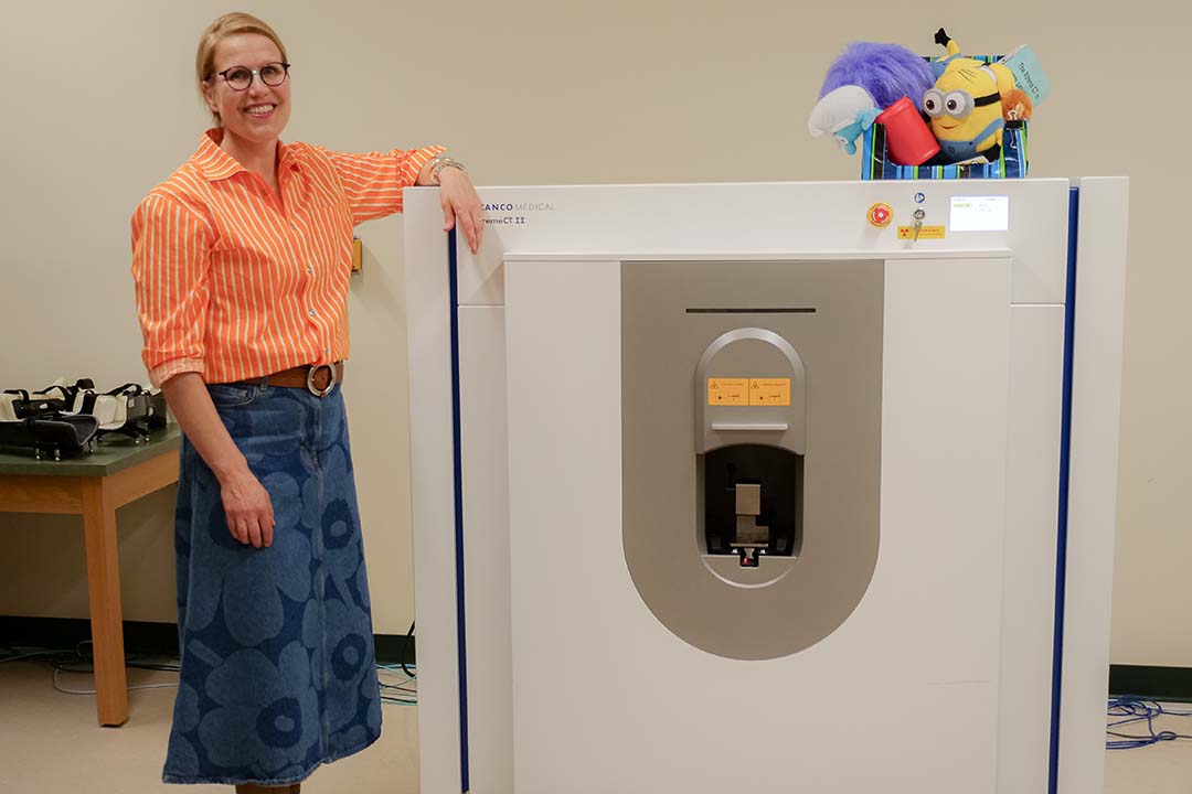
“The scanner is a second-generation, high-resolution peripheral quantitative computed tomography (HR-pQCT-II) and is a non-invasive way for us to look at the micro-architecture of the bone and the properties of the surrounding muscle,” said Kontulainen.
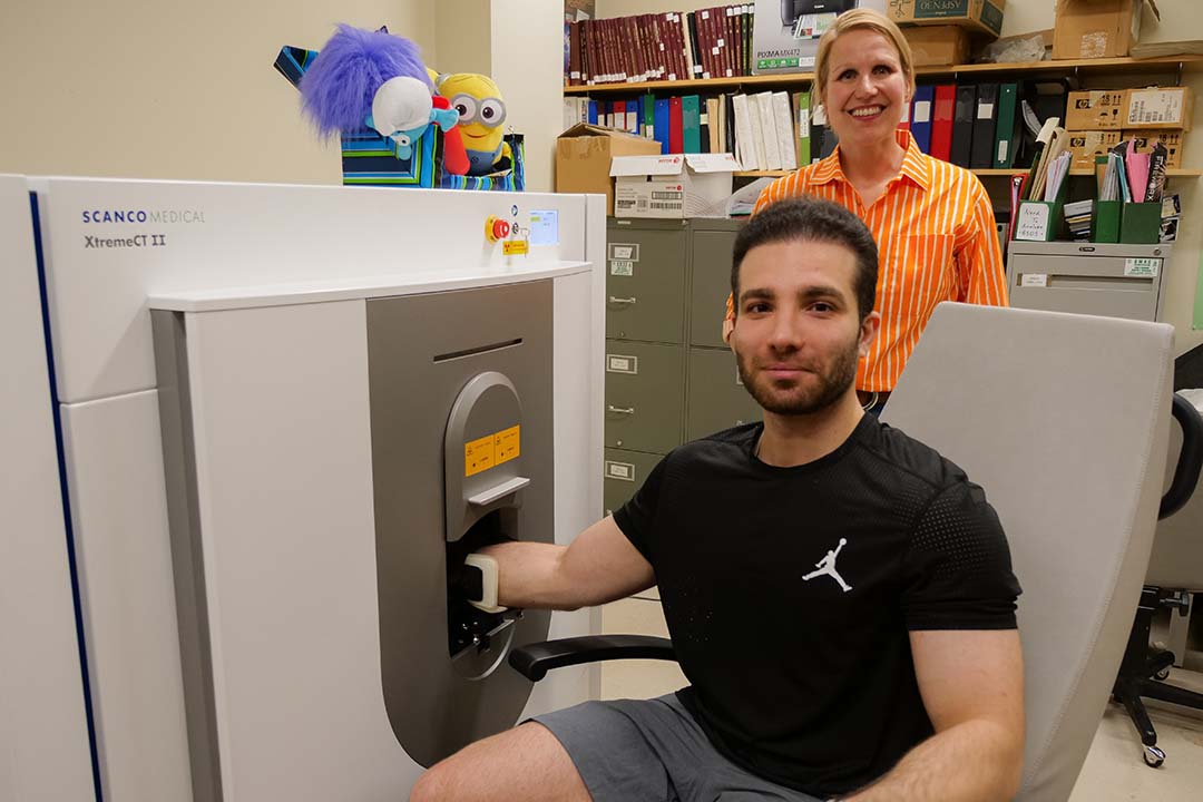
The powerful scanner is quite compact, taking up little space in the Laboratory for Imaging Muscle and Bone Structure (LIMBS) on the third floor of USask’s Physical Activity Complex. Children’s toys sit on top of the machine, used by the research team to keep the children calm and happy while they undergo a scan.
With detailed images of the inside of the bone and the surrounding structures, the team can run engineering simulations that pinpoint just how much force it will take to fracture the bone. These precise details help the team develop new evidence-based approaches for bone health assessments and develop therapies to monitor fracture prone populations, such as children with Type 1 diabetes and those diagnosed with autism spectrum disorder (ASD).
“These images are so important. We can use them to monitor how bone structure and strength develops, how it deteriorates and find out when changes begin in children who are prone to fractures,” said Kontulainen. “We also gain new insights into what influences bone development. This is all exciting because it offers monitoring of bone tissue in growing and aging skeleton, and the impact of therapies across the lifespan.”
Kontulainen said that bone health among children with Type 1 diabetes and children with ASD is often overlooked and poorly understood, but healthy bones are crucial for longevity. Being able to better understand bone development can help inform therapies, including non-pharmaceutical interventions like lifestyle modifications and physical activity. According to Kontulainen, early intervention can optimize bone strength and increase quality of life into older adulthood.
Kontulainen said that the new scanner is attracting interest from researchers and trainees across campus who need high-resolution images for their clinical and foundational studies.
“This scanner can be used for so many research projects and clinical studies across all ages, across campus,” said Kontulainen. “Having these kinds of advanced research tools available for use at the university benefits many populations, researchers and trainees to collaborate, including national and international studies. That is why continued investment into research infrastructure like this is so important and impactful.”