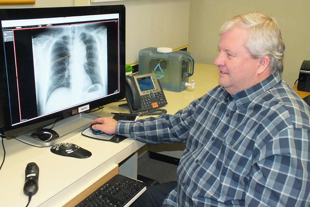
USask researcher creates new medical imaging technology
SASKATOON – As health care has evolved, medical imaging such as X-rays, CT scans, ultrasounds and MRIs have become increasingly important for diagnosis and treatment of patients. Through these imaging studies, health care professionals can learn how diseases are manifest to improve patient care.
A University of Saskatchewan (USask) research team has created unprecedented new solutions that enable medical students and other health care professionals to access high-quality diagnostic images and clinical information for teaching and learning—while protecting patient confidentiality.
“The way we manage images and use them for teaching has changed,” said Dr. Brent Burbridge of the USask College of Medicine’s Medical Imaging Department.
A big reason for this has been that digital images have replaced the traditional film images that were displayed on lightboxes. Today, digital images can be accessed on laptops, tablets
There have been downsides to the migration from physical to digital images. First, all confidential patient information has to be removed before the images can be used to teach students and residents. And second, there were problems with the portability and quality of the images available for teaching.
That is where Burbridge and his team come in.
They have developed software that enables images to be stored in a database with information such as patient age, gender
Next, they created an image viewing tool called ODIN—a repository of high-quality images, that can be annotated and downloaded, or reviewed in the online viewer, simulating a clinical workstation.
“One of the strengths of ODIN is its integrated search tool,” said Burbridge. “So, if you’re giving a lecture on tuberculosis, you type ‘tuberculosis’ into the search panel and all the cases we’ve created that having anything to do with tuberculosis come up. The teacher, or student, can review the cases and determine whether any of the images are useful for what they need.”
ODIN is short for Online DICOM Image Navigator. DICOM (Digital Imaging and Communications in Medicine) is the international standardized file format used for the creation, storage and communication of images in the clinical environment.
Until ODIN was developed, medical educators were only able to use lower quality JPEG copies of the DICOM images. These images had to be cropped or blacked out to remove identifiable patient information before use in teaching. Also, the ability to zoom in on the JPEG images without a loss of resolution limited their usefulness in some situations.
There is
Burbridge’s work was recently published in Medical Science Educator, a U.S.-based journal that focuses on teaching sciences which are fundamental to modern medicine and health.
To ensure that the technology continues to evolve and grow, Burbridge is working with I Innovation Enterprise (IE) to raise awareness of the technology and explore potential industrial partnerships and out-licensing opportunities. IE is the USask unit charged with maximizing the impact of knowledge-intensive innovations developed by researchers.
“We want to push forward,” said Burbridge. “We want to enhance what we’re doing—to improve it. Like any software, it needs refreshing. It needs new functionality.”
The research was made possible with funding provided by SaskTel through the Royal University Hospital Foundation, and the USask College of Medicine.
-30-
For more information, contact:
Jennifer Thoma
Media Relations Specialist
University of Saskatchewan
306-966-1851
jennifer.thoma@usask.ca

