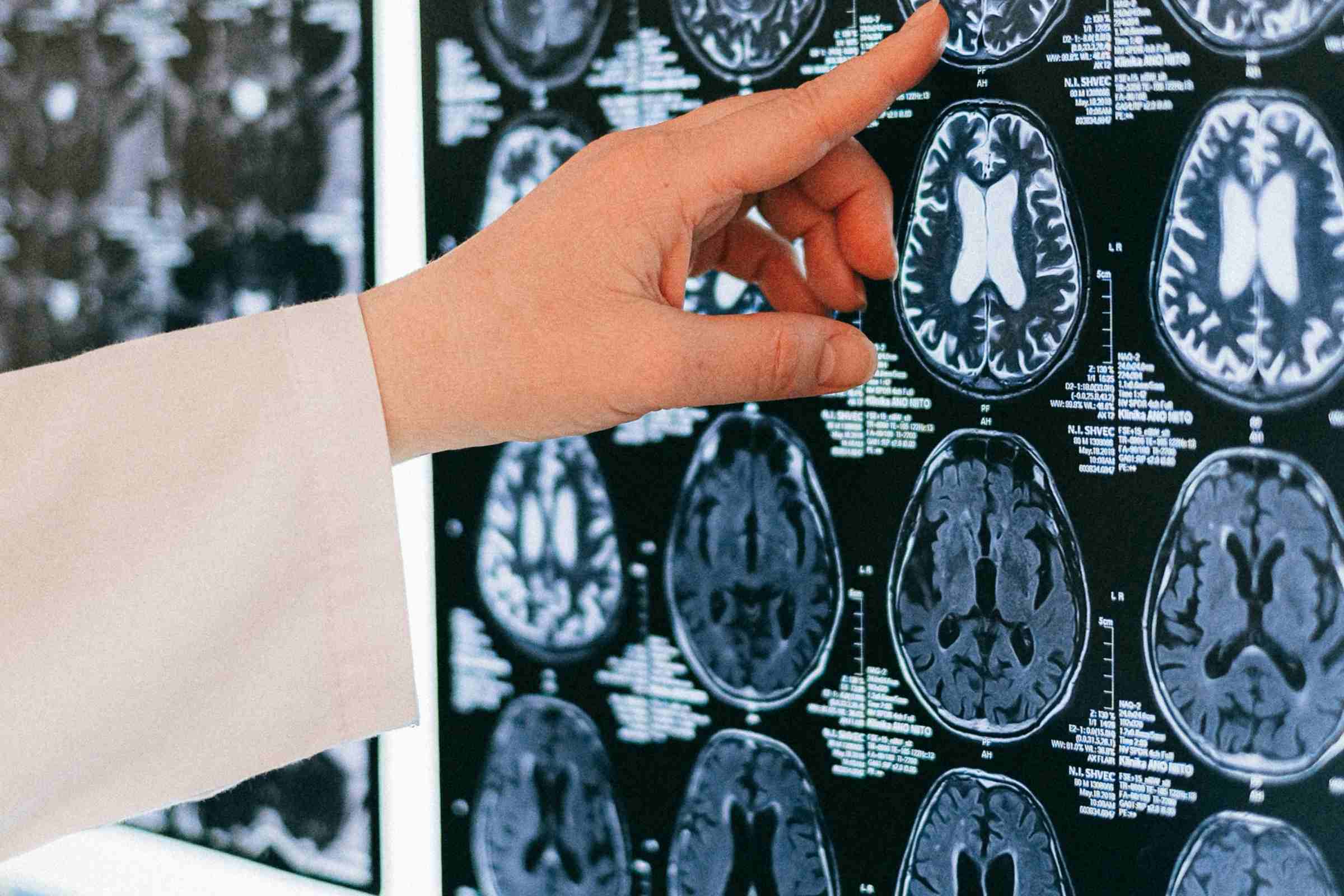
Brain differences in men and women could affect post-stroke outcomes
Strokes are the third leading cause of death in Canada and have impacted close to 880,000 Canadians over the age of 20.
By Erin Matthews“A stroke is caused by a loss of blood flow to brain cells. Without urgent intervention this may cause those cells to die because they constantly need energy and nutrients from the blood,” said Nicole Sylvain, clinical research coordinator and lab manager for Dr. Michael Kelly, a stroke neurosurgeon and professor who’s part of the Saskatchewan Cerebrovascular Center at the University of Saskatchewan
Sylvain and her colleagues are looking at treatments for post-stroke recovery that help supplement these energy losses. Using the Canadian Light Source (CLS) at the University of Saskatchewan (USask), the team was able to identify energy biomarkers in the brain — additional data that could eventually provide clinicians with critical information about the effects of potential stroke treatments on brain recovery after a stroke.
The group’s most recent paper published in IBRO Neuroscience Reports looked at differences post-stroke between male and female mice, and found that female mice have higher amounts of glycogen in their brains. Glycogen is a sugar-like substance that circulates in our blood and nourishes our cells. When the supply of glycogen is disrupted by stroke, the brain is severely impacted.
While men and women are almost equally affected by stroke, women tend to have poorer health outcomes. Animal models in stroke research traditionally use male subjects, but Sylvain and the team has identified a potential problem with taking the data from these lab experiments into clinical trials.
“We found that, for the most part, male data can be generalized for females, however, some of the metabolic markers we measured were actually different,” Sylvain said. “It’s really important to do the research on both sexes.”
It would be impossible for the team to detect the biomarkers without to the Mid-IR beamline.
“The only way to detect them in such an accurate way across the brain is with infrared imaging, so the CLS has been absolutely vital to our research.”
Newton, J. M., M. J. Pushie, N. J. Sylvain, H. Hou, S. Weese Maley, and M. E. Kelly. "Sex differences in the mouse photothrombotic stroke model investigated with X-ray fluorescence microscopy and Fourier transform infrared spectroscopic imaging." IBRO neuroscience reports 13 (2022): 127-135. doi.org/10.1016/j.ibneur.2022.07.006
Photos: Synchrotron | Mid-IR beamline | Nicole Sylvain
Article re-posted on .
View original article.

